Hip Arthroscopy — What It Is, Who Needs It, and What Recovery Looks Like

anatomical model of a hip bone.
The hip gets less arthroscopic attention than the knee or shoulder — it is a deeper joint, more technically demanding to access, and the range of conditions that can be treated inside it has only become well-defined over the past two decades. But for the right patients, hip arthroscopy is genuinely transformative: it resolves pain that has resisted months of conservative treatment and returns people to active lives without the recovery demand of open hip surgery.
The challenge is that hip pain has many causes, not all of them inside the joint. Getting the right diagnosis — determining that the pain is genuinely coming from a treatable intra-articular problem — is the most important step before any discussion of surgery.
This article explains what hip arthroscopy treats, who is a good candidate, what the procedure involves, and what recovery looks like.
What Hip Arthroscopy Can Reach
The hip is a ball-and-socket joint. The ball is the femoral head (top of the thigh bone). The socket is the acetabulum (part of the pelvis). The joint is surrounded by a capsule and stabilised by the labrum — a ring of fibrocartilage that deepens the socket and seals the joint.
An arthroscope inserted into the hip joint allows the surgeon to visualise:
- The acetabular labrum (the fibrocartilage rim)
- The articular cartilage of both the femoral head and acetabulum
- The ligamentum teres (a ligament inside the joint)
- The synovial lining of the joint
- The bony shape of the femoral neck and acetabular rim (from outside the joint through the peritrochanteric space)
Instruments inserted through separate portals can trim, repair, or reshape these structures.
Conditions Treated with Hip Arthroscopy
1. Femoroacetabular Impingement (FAI)
FAI is the most common condition treated by hip arthroscopy. It occurs when the ball and socket do not fit together smoothly — either because of abnormal bone growth on the femoral head (cam morphology), on the acetabular rim (pincer morphology), or both.
With every movement of the hip, this abnormal bone shape causes the femoral head to impinge against the acetabular rim, damaging the labrum and, over time, the cartilage. The result: groin pain with activity, particularly hip flexion — squatting, getting in and out of cars, cycling, running.
Arthroscopic treatment removes the excess bone (femoroplasty or acetabuloplasty) and repairs any labral damage. Studies consistently show excellent outcomes for FAI in well-selected patients — most return to full activity, including sport, within 4–6 months.
2. Hip Labral Tears
The labrum can tear from trauma, from FAI (which causes repeated impingement stress on the labrum), or from structural abnormalities like hip dysplasia (a shallow socket).
Labral repair — reattaching the torn labrum to the acetabular rim with small suture anchors — is preferable to labral debridement (trimming the torn portion). Preserving labral function maintains joint stability and reduces the risk of progressive cartilage damage.
Labral repair outcomes are better when the underlying cause (e.g., FAI) is also addressed at the same time. Repairing the labrum without correcting the bony impingement that caused the tear often leads to recurrence.
3. Loose Bodies
Fragments of cartilage or bone floating inside the hip joint cause sharp, intermittent pain, clicking, and catching. Hip arthroscopy removes these loose bodies efficiently.
4. Synovitis and Inflammatory Conditions
Inflammation of the hip joint lining — from inflammatory arthritis, infection, or unexplained synovitis — can be managed arthroscopically through synovectomy (removing inflamed tissue) and joint washout.
5. Hip Dysplasia (Mild Cases)
In mild hip dysplasia — where the acetabulum is shallower than normal but not severely so — arthroscopy can address the labral and cartilage consequences. Severe dysplasia requires periacetabular osteotomy (PAO), a larger reconstructive procedure that is outside the scope of arthroscopy.
Who Is the Right Candidate?
Hip arthroscopy works best in
Younger patients (under 45–50) without significant arthritis. The procedure is designed to address structural problems — impingement, labral tears, loose bodies — in a joint that still has good cartilage. In advanced osteoarthritis, where the cartilage is significantly worn, the underlying disease process will continue regardless of what is done arthroscopically.
Patients with confirmed intra-articular pathology on imaging. An MRI with arthrogram (where dye is injected into the joint) is often the best imaging for hip labral tears and cartilage damage. Plain MRI can miss labral pathology. The imaging must show something specific that can be treated.
Patients who have had adequate conservative management. Hip pain from FAI or labral tears should be given a trial of physiotherapy — hip strengthening, core stability, and activity modification — before surgery is considered. Many patients with mild FAI manage well without surgery.
Patients without advanced hip dysplasia. Shallow sockets are a contraindication to standard hip arthroscopy without concurrent PAO — operating on a dysplastic hip without addressing the dysplasia leads to poor outcomes.
Hip Arthroscopy Recovery
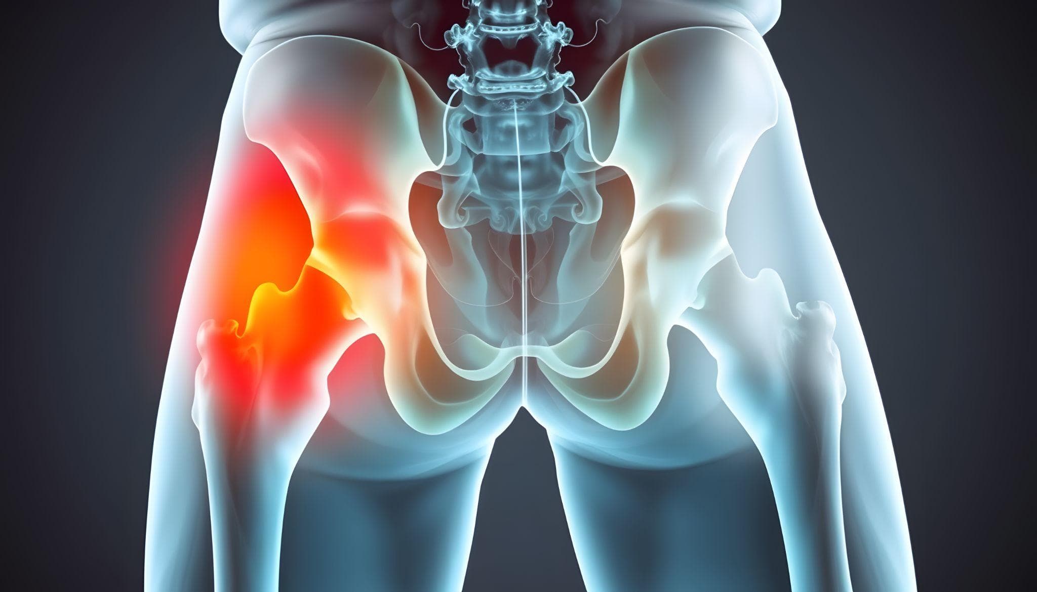
X-ray style illustration highlighting pain and damage in the hip joint.
The hip is a weight-bearing joint. Recovery after hip arthroscopy requires a balance between protecting the repair and preventing the stiffness and muscle wasting that come from too much rest.
Day 0–2: Day surgery. Crutches from day one. Ice and elevation. Physiotherapy begins immediately with isometric exercises (tensing without movement) and gentle range of motion.
Week 1–4 (for labral repair / FAI):
- Partial weight-bearing with crutches
- No hip flexion beyond 90 degrees
- Passive hip stretching is restricted to protect the repair
- Aquatherapy (pool walking) introduced early when incisions are healed
Week 4–8:
- Full weight-bearing — crutches phased out
- Progressive physiotherapy: hip abductor, flexor, and core strengthening
- Stationary cycling starts
Month 2–4:
- Walking confidently without aids
- Sport-specific rehabilitation begins
- Running is introduced progressively from month 3–4
Month 4–6:
- Return to full sport for most patients with labral repair and FAI
- Athletes in high-demand sports (football, martial arts, basketball) may require 6–9 months
For simple procedures (loose body removal, synovectomy without labral repair), recovery is faster — most patients are walking normally within 2–3 weeks.
What Hip Arthroscopy Cannot Do
It is important to be clear: hip arthroscopy is not a substitute for hip replacement in advanced arthritis. If the cartilage is gone, impingement correction and labral repair will not produce good outcomes — the underlying damage is already irreversible.
Similarly, severe hip dysplasia requires pelvic osteotomy, not arthroscopy. Operating arthroscopically on a severely dysplastic hip without correcting the dysplasia exposes the labrum and cartilage to continued abnormal loading.
The patient selection conversation — establishing whether a patient's hip is a good arthroscopy candidate — is as important as the surgery itself.
Frequently Asked Questions
Q: How is hip arthroscopy different from hip replacement?
Hip arthroscopy is minimally invasive and addresses specific problems inside an otherwise healthy or mildly damaged joint. Hip replacement removes and replaces the entire joint surface. They are not comparable procedures — arthroscopy is for patients with structural problems in a relatively good joint; replacement is for end-stage disease.
Q: Will I have a scar after hip arthroscopy?
The portals are each about 5mm — the scars are minimal and fade significantly over months.
Q: Is hip arthroscopy available in Greater Noida?
Yes. Dr. Akash Dubey holds a Fellowship in Arthroscopy and Sports Surgery and performs hip arthroscopy at KDSG Superspeciality Hospital, Greater Noida.
Q: Can I return to running after hip arthroscopy?
Yes, for most patients. Running typically resumes at 3–4 months post-operatively after FAI correction and labral repair, with progression to full sport at 5–6 months.
Q: What happens if hip arthroscopy does not work?
In patients where symptoms persist despite technically successful arthroscopy, the next steps are reassessed — including whether an additional structural cause was missed, or whether the degree of underlying cartilage damage was more significant than pre-operative imaging suggested.
Contact Dr. Akash Dubey — Your Bone Mechanic
Dr. Akash Dubey
MBBS (KGMU, Lucknow) | MS | DNB Ortho | FIAS | FIJR | FIFA Diploma in Football Medicine
Robotic Surgery Certified | Member, AOPAS
Clinic: Shop No. 24, Ground Floor, Gaur City Arcade, Near Sarvodaya Hospital, Gaur City 2, Greater Noida West, U.P. – 201301
To book a consultation for hip arthroscopy in Greater Noida, call +91-8130441429